Albugo candida (Cystopus): Life Cycle, Reproduction & White Rust
Comprehensive study notes on Albugo candida (Cystopus): covering general features, systematic position, asexual and sexual reproduction, life cycle diagram, and white rust disease control for BSc Botany students.

Albugo candida (Cystopus): General Features, Reproduction, Life Cycle and White Rust Disease
Introduction to Albugo candida (Cystopus)
Albugo candida (Cystopus) is an obligate parasitic oomycete that causes white rust, also called white blister rust, on mustard, rapeseed, radish, cabbage, cauliflower and many other members of the family Brassicaceae (Cruciferae). It is a standard type study in BSc Botany because it shows, in a single organism, a coenocytic mycelium, haustoria, zoosporic asexual reproduction, oogamous sexual reproduction and a well-defined disease cycle. These notes cover the syllabus topics in order, from general features and systematic position to life cycle, white rust symptoms and control measures.
Common Name and Scientific Name
- Common names: white rust, white blister rust, white blister, staghead disease (for the systemic flower-head phase).
- Accepted scientific name: Albugo candida (Pers.) Kuntze.
- Older name still used in many textbooks: Cystopus candidus (Pers.) Lév. Another synonym is Albugo cruciferarum (DC.) Gray. Because the older name appears in many university syllabi, this note uses "Albugo (Cystopus)" wherever helpful.
Why Is Albugo candida Studied in BSc Botany?
- It is a representative member of the Oomycota, a group of fungus-like organisms that are not true fungi. Studying it teaches students how to separate look-alike groups.
- It shows a complete oogamous life cycle with distinct male (antheridium) and female (oogonium) gametangia and a thick-walled resting oospore.
- It is a classic example of obligate parasitism, with intercellular hyphae and intracellular haustoria.
- It links lower-plant studies with plant pathology, because the same organism is a serious pathogen of oilseed and vegetable crops.
Importance as a Plant Pathogen
Albugo candida occurs wherever crucifers are grown and can destroy leaves, stems and inflorescences. In Indian mustard, white rust is regarded as one of the most important diseases and is commonly reported to reduce seed yield by roughly 17–34%, with much higher losses recorded in severe epidemics. The pathogen is also a valuable research organism: it is used to study how obligate biotrophs obtain nutrients and suppress plant defences.
Brief Introduction to White Rust Disease
Although the disease is called a "rust", it has nothing to do with the true rusts (order Pucciniales, Basidiomycota), which are caused by fungi such as Puccinia. The name arises only because the pustules break through the epidermis in a rust-like manner.
Table of Contents
General Features of Albugo candida
General Nature and Biological Characteristics
Albugo candida is a eukaryotic, non-photosynthetic, heterotrophic organism that lives as an obligate biotrophic parasite inside the living tissues of crucifers. It lacks chlorophyll and feeds by absorbing dissolved nutrients from living host cells. It has a filamentous, fungus-like body and was therefore placed for a long time among the "lower fungi" (the old group Phycomycetes). Modern research shows that it belongs to a separate evolutionary lineage, the Stramenopiles, and is closer to brown algae and diatoms than to mushrooms or moulds.
Classification as an Oomycete
The word oomycete means "egg fungus" (Greek oon, egg; mykes, fungus). It refers to the characteristic sexual process in which a large, non-motile egg cell inside an oogonium is fertilised to form a thick-walled oospore. Albugo candida belongs to the Oomycota and, within it, to the order Albuginales (see the next section).
Thallus Organisation
The thallus is a well-developed, profusely branched mycelium made of coenocytic (aseptate) hyphae that grow between the host cells (intercellular). Small, knob-like haustoria penetrate host cells. The thallus is eucarpic: only a part of it becomes reproductive (sporangiophores, gametangia) while the rest continues as vegetative mycelium.
Nutrition
Nutrition is heterotrophic and biotrophic. The pathogen absorbs sugars, amino acids and other nutrients through the haustoria without killing the host cell quickly. It cannot be grown on ordinary artificial culture media, which is a defining feature of an obligate parasite.
Parasitic Habit
The pathogen may cause local infection (limited pustules on leaves and stems) or systemic infection (mycelium spreading through the stem and inflorescence, producing stagheads).
Cell Wall Composition
The hyphal wall consists mainly of cellulose and β-glucans, together with hydroxyproline-rich protein. Chitin, the major structural polysaccharide of true fungal walls, is absent or present only in trace amounts. This is one of the reasons why oomycetes are not classified with true fungi.
Reproduction
- Asexual: by sporangia (called conidia in many older textbooks) borne in chains on sporangiophores; they usually germinate to release biflagellate zoospores.
- Sexual: oogamous, by contact between an antheridium and an oogonium, giving a resistant oospore.
- Vegetative reproduction by fragmentation is mentioned in some textbooks but is of minor importance (see the reproduction section).
Important Distinguishing Features
- Obligate parasite of Brassicaceae producing white, chalky pustules.
- Coenocytic, intercellular mycelium with knob-like haustoria.
- Sporangiophores are club-shaped and form a compact palisade layer below the host epidermis.
- Sporangia are formed in basipetal chains with disjunctors (gelatinous connecting pads) between them.
- Zoospores are biflagellate and heterokont (one tinsel flagellum and one whiplash flagellum).
- Oogonium has a single, uninucleate oosphere with a central coenocentrum; the antheridium is club-shaped and paragynous.
- Oospore has a thick, brown, ornamented wall and acts as the resting spore.
Albugo candida Compared with True Fungi
| Feature | Albugo candida (Oomycota) | True fungi (Eumycota) |
|---|---|---|
| Evolutionary group | Stramenopiles (SAR clade); fungus-like protist | Kingdom Fungi (related to animals) |
| Cell wall | Cellulose and β-glucans; chitin absent or only traces | Chitin with glucans |
| Nuclear state of vegetative body | Diploid (2n) | Usually haploid or dikaryotic |
| Hyphae | Coenocytic (aseptate); septa only to cut off reproductive structures or in old or injured hyphae | Aseptate in some groups; regularly septate in Ascomycota and Basidiomycota |
| Motile spores | Biflagellate, heterokont zoospores | Absent in most groups; posterior single flagellum in chytrids |
| Sexual spore | Oospore (oogamy) | Zygospore, ascospore or basidiospore |
| Reserve food | Mycolaminarin (a β-glucan) and lipids | Glycogen and lipids |
| Sterols | Most oomycetes cannot synthesise sterols and depend on the host | Synthesise ergosterol |
| Fungicide response | Sensitive to anti-oomycete chemicals such as phenylamides; many azole fungicides are generally poorly effective | Azoles are widely effective |
Systematic Position of Albugo candida
The classification of oomycetes has changed greatly. Traditional textbooks place Albugo in the Kingdom Fungi, whereas modern phylogenetic classifications place it among the Stramenopiles, outside the true fungi. Both schemes are given below so that students can answer according to the classification prescribed by their university.
Modern (Molecular) Classification
| Rank | Taxon |
|---|---|
| Domain | Eukaryota |
| Supergroup / Clade | SAR → Stramenopiles (Straminipila) |
| Kingdom (in some schemes) | Chromista |
| Phylum / Division | Oomycota (= Peronosporomycota in some schemes) |
| Class | Peronosporomycetes (= Oomycetes / Peronosporea) |
| Order | Albuginales |
| Family | Albuginaceae |
| Genus | Albugo |
| Species | Albugo candida (Pers.) Kuntze |
Traditional Textbook Classification
| Rank | Taxon |
|---|---|
| Kingdom | Fungi (Mycota) |
| Division | Eumycota |
| Sub-division | Mastigomycotina |
| Class | Oomycetes |
| Order | Peronosporales |
| Family | Albuginaceae |
| Genus | Albugo (Cystopus) |
| Species | candida (candidus) |
Why Do the Two Classifications Differ?
- Kingdom and phylum: Early mycologists grouped all filamentous, spore-producing, non-photosynthetic organisms together as fungi. Studies of cell wall chemistry, flagella, biochemistry and gene sequences later showed that oomycetes are Stramenopiles. The kingdom name for this lineage varies among systems (for example Chromista in Cavalier-Smith's system), and many modern schemes use clade names without a kingdom rank.
- Order: Albugo and its relatives were long kept in the Peronosporales together with the downy mildews. Molecular and morphological evidence showed that white rusts form a separate lineage, and they are now placed in their own order, Albuginales. Older books still use Peronosporales.
- Sub-division: The sub-division Mastigomycotina (zoosporic fungi) is used only in traditional systems.
Habit and Habitat of Albugo candida
Type of Organism and Parasitic Nature
Albugo candida is an obligate parasite (biotroph). Its hyphae live inside the host, and only the sporangial pustules and, at a later stage, the gametangia and oospores are found in association with the host tissue. There is no free-living, saprophytic phase.
Host Plants
The principal hosts belong to the family Brassicaceae (Cruciferae). Older accounts list additional families, but many of those records refer to other Albugo species or to species now placed in other genera.
| Group | Representative hosts |
|---|---|
| Oilseed crops | Indian mustard (Brassica juncea), rapeseed / canola (B. napus), turnip rape (B. rapa) |
| Vegetable crops | Cabbage and cauliflower (B. oleracea), radish (Raphanus sativus), rocket / arugula (Eruca vesicaria), horseradish (Armoracia rusticana) |
| Wild and weedy crucifers | Shepherd's purse (Capsella bursa-pastoris) and various wild mustards, which act as reservoirs of inoculum |
| Model plant | Arabidopsis thaliana (used in laboratory studies of white rust interactions) |
Different isolates are often named races or pathotypes after the host from which they were first obtained (for example, isolates from B. juncea or R. sativus). Their host ranges overlap, but each race is typically most aggressive on its original host.
Suitable Environmental Conditions
- Temperature: Cool weather favours infection. Infection and zoospore activity occur over a range of roughly 10–25 °C, with the best conditions in the cooler part of that range. Hot, dry weather checks the disease.
- Moisture: Free water on the leaf surface (dew, fog, rain, sprinkler irrigation) and high humidity are needed because the zoospores swim in water films.
- Season: In South Asia the disease is mainly a winter (rabi) season problem of mustard. In temperate regions it is prominent in cool, moist spring and autumn weather.
Geographical Distribution
The pathogen is cosmopolitan. It has been reported from Europe, Asia, Africa, Australasia and the Americas, wherever crucifers are cultivated. It is especially important in the major rapeseed–mustard regions of India, China, Canada, Australia and Europe.
Occurrence on Agricultural and Wild Plants
On crops, it attacks seedlings, mature leaves, stems, flowers and pods. On wild crucifers, it persists between crop seasons and provides sporangia that start new infections in nearby fields.
Relationship Between the Pathogen and the Host
The relationship is one of biotrophy. The mycelium grows in the intercellular spaces and sends haustoria into living cells. The host cells remain alive, but their metabolism is redirected to feed the pathogen. The parasite also secretes proteins called effectors that weaken host defence responses and disturb normal growth, which explains the swelling and distortion seen in infected tissues.
Somatic Structure of Albugo candida
The somatic (vegetative) body of Albugo candida is a mycelium that lives entirely inside the host plant. It is studied under the light microscope in stained sections of infected leaves or stems, where the hyphae are seen running between the host cells with small haustoria inside them.
Structure of the Vegetative Thallus
The thallus consists of a network of fine, colourless, much-branched hyphae. Together these hyphae form a eucarpic, intercellular mycelium that spreads through the spongy mesophyll of leaves, and through the cortex and pith of stems and flower stalks. No part of the vegetative mycelium grows on the outside of the plant.
Hyphal Organisation and Branching
The hyphae are irregularly and profusely branched. They follow the intercellular spaces, squeezing between host cells and passing around them. Branching increases the surface area in contact with host cells and allows the pathogen to occupy large volumes of tissue. Beneath the epidermis the hyphae become densely packed and give rise to the reproductive structures.
Septation: The Coenocytic Condition
The hyphae are aseptate, that is, they lack regular cross-walls. Such a hypha is called coenocytic: a single, continuous, tubular mass of cytoplasm that contains many nuclei.
Significance of the coenocytic condition:
- Cytoplasm, nutrients and nuclei move freely along the hypha, supporting rapid growth and rapid development of reproductive structures.
- Growth is concentrated at the hyphal tips, which advance quickly through the host tissue.
- Septa do form in a few situations: to cut off sporangia, gametangia and other reproductive structures, and to seal off old or damaged parts of a hypha.
Hyphal Wall
The wall is thin and made largely of cellulose and β-glucans. Chitin is absent or occurs in traces only. This chemistry is different from that of true fungi.
Cytoplasm and Nuclei
The cytoplasm is dense and granular and contains numerous small diploid nuclei scattered along the hypha, mitochondria with tubular cristae, endoplasmic reticulum, Golgi bodies, vacuoles and oil (lipid) droplets as food reserves. Because the vegetative nuclei are diploid, Albugo is described as a diplontic organism.
Haustoria
From the intercellular hyphae, short lateral branches press against the wall of a host cell and penetrate it. Inside the cell, the tip enlarges into a small haustorium. In Albugo candida haustoria are typically small, unbranched, knob-like or globose and are attached to the hypha by a narrow neck. The haustorium does not burst through the host plasma membrane; instead it pushes the membrane inwards, so it remains separated from the host cytoplasm by the host membrane and a thin extrahaustorial matrix.
Function of Haustoria
- Absorption: They take up sugars, amino acids and other nutrients from the living host cell.
- Feeding without killing: Because the host membrane remains intact, the cell stays alive, which is the essence of biotrophy.
- Secretion of effectors: Modern studies show that haustoria are also sites where the pathogen delivers effector proteins that suppress host immunity.
Relationship Between the Pathogen and the Host Cells
The pathogen lives in the intercellular spaces and connects with living cells only through haustoria. Infected tissues show hypertrophy (abnormal enlargement of cells) and hyperplasia (abnormal increase in cell number), which produce swellings and distortion. Disturbance of the host's growth regulation is believed to contribute to these changes.
Internal and External Growth
- Internal growth: All vegetative growth is internal and intercellular. It may be localised near the point of entry or systemic along stems, petioles and inflorescences.
- External appearance: The pathogen becomes visible only when the sporangial masses push up and rupture the host epidermis to form white pustules.
Reproduction in Albugo candida
Albugo candida reproduces by three methods: vegetative, asexual and sexual. Of these, asexual reproduction spreads the disease during the crop season, whereas sexual reproduction produces the resting oospores that carry the pathogen through unfavourable periods.
A. Vegetative Reproduction
Meaning
Vegetative reproduction is the formation of new individuals from a part of the vegetative body without the involvement of spores or gametes.
Fragmentation and Its Process
Some textbooks state that the mycelium of Albugo may reproduce by fragmentation. In this process a hypha breaks into pieces, each with cytoplasm and nuclei. A fragment that remains inside living host tissue continues to grow, branch and form a new mycelium.
Formation of New Individuals
New individuals arise only if the fragment stays in contact with living host cells, because the pathogen cannot survive as a free-living saprophyte. Practically, this means that mycelium can persist and continue to grow in perennial or vegetatively propagated hosts and in living infected tissues.
Significance
- It allows mycelium to survive within perennial hosts between seasons.
- It contributes in a small way to the spread of the pathogen within an infected plant.
B. Asexual Reproduction
Meaning
Asexual reproduction is the production of new individuals through spores formed without the fusion of gametes. In Albugo the asexual spores are borne in chains at the tips of special hyphal branches and function mainly as zoosporangia.
Conditions Favouring Asexual Reproduction
- A mature, well-established mycelium in a healthy, actively growing host.
- Cool temperatures and high humidity.
- Abundant dew, fog or rain for the later germination and infection stages.
Formation of Sporangiophores
As the mycelium matures, hyphae accumulate just beneath the host epidermis (usually the lower epidermis of the leaf). Short, unbranched, club-shaped (clavate) sporangiophores arise from these hyphae and stand side by side in a compact layer resembling a palisade, oriented perpendicular to the surface. Together they form a sorus. Each sporangiophore is multinucleate and rich in dense cytoplasm.
Arrangement and Development of Sporangia (Conidia) in Chains
- The swollen tip of each sporangiophore becomes cut off by a septum, forming the first sporangium.
- The sporangiophore continues to form new sporangia below the first one by repeated abstriction (pinching off) at its tip.
- A chain is therefore produced in basipetal succession: the oldest sporangium is at the tip of the chain and the youngest is at the base, next to the sporangiophore.
- Between successive sporangia lies a small disjunctor, an intercalary gelatinous pad that holds neighbouring sporangia together and later helps them separate.
- Mature sporangia are spherical to slightly oval, hyaline (colourless) and thin-walled, and measure about 12–22 µm in diameter (commonly around 14–18 µm).
Liberation of Sporangia
The continued formation of sporangia builds up pressure beneath the epidermis. The epidermis first bulges into a blister and then ruptures, exposing a white, powdery mass of sporangia. The disjunctors dry or dissolve, and the sporangia become separate. They are dispersed by wind, rain splash and insects, and may also be carried on tools and machinery.
Formation and Liberation of Zoospores
Sporangia that land on a moist leaf of a suitable host in cool conditions usually germinate indirectly:
- The multinucleate protoplasm of the sporangium divides into a small number of uninucleate portions, commonly about 4–8 (often 5–7).
- Each portion differentiates into a biflagellate, kidney-shaped zoospore with two unequal flagella of the heterokont type: one forward-directed tinsel flagellum and one backward-directed whiplash flagellum.
- The sporangial wall opens (often through a pore, with the contents passing into a thin vesicle), and the zoospores are released.
- The zoospores swim for a short time in the film of water on the leaf surface.
Some accounts also report direct germination of sporangia by a germ tube, especially under less favourable conditions. Zoospore formation is the usual and best-documented route.
Germination of Spores and Infection of Host Plants
- Zoospores are attracted to the host surface, especially to stomata. They encyst (lose their flagella and secrete a wall) near a stoma.
- Each cyst produces a germ tube that grows through the stomatal opening into the sub-stomatal cavity.
- The germ tube develops into an intercellular mycelium, forms haustoria, and establishes the parasitic relationship.
- In experimental studies on mustard, the time from inoculation to the first symptoms (incubation period) and to production of new sporangia (latent period) is of the order of several days to about two weeks, depending on isolate, host stage and temperature.
Significance of Asexual Reproduction
- It produces enormous numbers of sporangia in a short time.
- It gives rise to secondary infections and repeated disease cycles during a single crop season, so that a small outbreak can become an epidemic.
- It allows rapid spread of the disease within and between fields by wind and rain.
C. Sexual Reproduction
1. Meaning of Sexual Reproduction
Sexual reproduction is the formation of a new individual after fusion of two compatible gametic nuclei (karyogamy), usually preceded by fusion of cytoplasm (plasmogamy). It combines the genetic material of two parents.
2. Oogamous Type of Sexual Reproduction
In Albugo candida sexual reproduction is oogamous: it involves a large, non-motile female gamete (the oosphere or egg) and a male nucleus delivered through a tube. It is gametangial contact: the male and female gametangia (antheridium and oogonium) meet and the male nucleus is transferred through a fertilization tube, without free-swimming gametes. The male gametangium does not release a motile sperm.
Sexual structures are formed mostly late in the season in the deeper tissues of stems, flower stalks and stagheads, and sometimes in leaves, in the intercellular spaces of the host.
3. Formation of Antheridia
The antheridia develop from short hyphal branches near the young oogonium. The tip of such a branch swells and is cut off by a septum. It is attached beside the oogonium, which is described as a paragynous arrangement (the antheridium is applied to the side of the oogonium rather than surrounding its base).
4. Formation of Oogonia
The oogonium begins as a terminal swelling of a hyphal branch. It enlarges, becomes rounded, and is separated from the supporting hypha by a septum. It contains dense multinucleate cytoplasm.
5. Structure of the Antheridium
The mature antheridium is club-shaped and multinucleate, with a thin wall. It lies against the oogonial wall and gives rise to a fertilization tube.
6. Structure of the Oogonium
The mature oogonium is globose (spherical), larger than the antheridium, and has a thick wall. Its contents become organised into:
- a peripheral layer of cytoplasm called the periplasm, and
- a dense central mass of cytoplasm called the ooplasm (the oosphere), inside which a specialised dense body called the coenocentrum appears.
Older cytological studies estimated that the young oogonium of A. candida contains many nuclei (of the order of 70–110). When the oosphere is differentiated, only one nucleus becomes functional as the female nucleus and lies near the coenocentrum. The other nuclei move into the periplasm and degenerate. The mature oosphere is therefore uninucleate. (In some other white rusts, the oosphere contains many functional nuclei, another reason why species must not be confused.)
7. Fertilization
- The antheridium comes into contact with the oogonium, and at the point of contact the oogonial wall may form a small, inconspicuous receptive papilla.
- The antheridium sends out a fertilization tube that penetrates the oogonial wall, grows through the periplasm and reaches the ooplasm.
- The tip of the tube opens and discharges one male nucleus (with a little cytoplasm) into the oosphere.
- The male nucleus approaches the female nucleus, and the two fuse (karyogamy) in the region of the coenocentrum, forming a diploid zygote nucleus.
8. Formation of the Oospore
After fertilization, the zygote (fertilised oosphere) secretes a thick wall around itself, using material from the ooplasm and periplasm. The zygote with its wall is the oospore.
9. Development and Maturation of the Oospore
- The oospore wall becomes three-layered: a thick, dark-brown outer exospore (epispore), a middle layer (mesospore) and a thin inner endospore.
- The exospore of A. candida is ornamented with ridges or tubercles.
- The cytoplasm becomes dense and rich in oil reserves, and the oospore enters dormancy.
- The oospores lie among the remains of the host tissue. As the stems and stagheads dry and decay, the oospores are released into the soil or remain in the debris. They may also contaminate seed lots as fragments of stagheads.
10. Germination of the Oospore
- After a period of dormancy, and when there is moisture and a suitable temperature, the oospore germinates.
- The thick exospore cracks, and the inner endospore protrudes as a thin-walled vesicle.
- The diploid protoplasm in the vesicle divides into many biflagellate zoospores. The number is variable, and textbooks quote figures from a few dozen (often 32–64) up to about 60.
- The vesicle bursts and the zoospores are released. They swim, encyst on a seedling of a crucifer, and infect it through a stoma (primary infection).
- Some accounts also mention germination by a germ tube. Oospore germination is difficult to observe under natural conditions, so accounts vary in detail.
11. Importance of Sexual Reproduction
- The oospore is a resting spore: it survives heat, drought, cold and the absence of a host.
- Oospores in soil, plant debris and seed lots are the main source of primary infection in the next season.
- Sexual reproduction allows genetic recombination, which can generate new races of the pathogen that overcome host resistance.
- It helps in long-distance spread of the pathogen through contaminated seed.
Life Cycle of Albugo candida
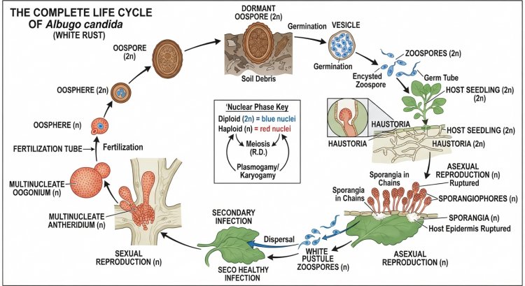
The life cycle of Albugo candida is diplontic: the vegetative mycelium and the spores derived from it are diploid, and the haploid phase is confined to the gametic nuclei formed just before fertilization. The life cycle is an autoecious (single-host) cycle completed entirely on crucifers. It consists of three linked phases: vegetative, asexual and sexual.
Vegetative Phase
After infection, the germ tube develops into an intercellular, coenocytic mycelium with haustoria. The mycelium branches through the host tissue and absorbs nutrients from living cells. It may remain local or become systemic.
Asexual Phase
During the growing season, mycelium beneath the epidermis gives rise to club-shaped sporangiophores. Chains of sporangia (conidia) are cut off in basipetal succession, rupture the epidermis and are dispersed. In water they release zoospores that initiate new infections. This phase is repeated many times in one season.
Sexual Phase
Late in the season, and mostly in stems, flower stalks and stagheads, oogonia and antheridia develop. A male nucleus is transferred through a fertilization tube to the oosphere, and a thick-walled oospore is formed.
Formation of Oospores, Survival and Germination
Oospores are released when the infected tissue decays. They survive in soil, in crop residues and in staghead fragments that contaminate seed. After dormancy, oospores germinate in moist conditions by forming a vesicle from which zoospores are released.
Primary Infection
Primary infection is the first infection of a crop in a season. Its sources are:
- Oospores in soil and infected crop debris, and oospore-containing staghead fragments mixed with seed.
- Perennating mycelium in perennial or volunteer host plants.
- Sporangia blown from infected volunteer crucifers, weeds and neighbouring crops.
Seedlings are infected through the cotyledons and young leaves.
Secondary Infection
Secondary infection is caused by sporangia produced in the pustules that arise from primary infection. These sporangia spread the disease from plant to plant and field to field, and each cycle takes only about one to two weeks under favourable conditions. Because the disease can go through many such cycles in one season, it is called polycyclic.
Repetition of the Disease Cycle
The disease cycle is repeated every crop season: oospores and other perennating inoculum start the epidemic, sporangia multiply it, and new oospores in the diseased stems and stagheads carry it over to the next season.
Relationship Between the Life Cycle and White Rust Disease
| Stage in life cycle | Corresponding event in white rust disease |
|---|---|
| Oospore germination and zoospores | Primary infection of seedlings |
| Vegetative mycelium with haustoria | Incubation period; chlorotic spots; nutrient drain |
| Sporangiophores and sporangia | White pustules on leaves, stems and pods |
| Zoospores from sporangia | Secondary infection and spread of the epidemic |
| Systemic mycelium | Stagheads, sterile flowers, distorted tissue |
| Oogonia, antheridia and oospores | Brown resting spores inside dead stems; source of next season's disease |
Diagrammatic Representation of the Life Cycle of Albugo candida
The text diagram below shows the cycle in the correct biological sequence. The linear order often given in syllabi (mycelium → asexual reproduction → infection → sexual reproduction → oospore → germination) has been corrected: the sexual phase does not follow directly after infection, but arises late in the season from the same mycelium, after several asexual cycles. The oospore germination is what starts the primary infection of the next season.
                        DORMANT OOSPORE
     (in soil, host debris, stagheads, or as seed contaminant)
                              │
                              │  moisture + cool temperature
                              ▼
            OOSPORE GERMINATION (exospore cracks)
                 endospore forms a vesicle
                              │
                              ▼
               ZOOSPORES (biflagellate, released)
                              │
                              ▼
     Encystment on seedling → germ tube → entry through stoma
                     (PRIMARY INFECTION)
                              │
                              ▼
                   VEGETATIVE MYCELIUM
     (intercellular, coenocytic, diploid, with haustoria)
                              │
            ┌─────────────────┴──────────────────┐
            ▼                                    ▼
   ASEXUAL REPRODUCTION                 SEXUAL REPRODUCTION
   (repeated through the season)        (late season; stems / stagheads)
            │                                    │
            ▼                                    ▼
   Club-shaped sporangiophores          Oogonium (globose; one oosphere
   below the epidermis                  with coenocentrum)
            │                            + Antheridium (club-shaped)
            ▼                                    │
   SPORANGIA (CONIDIA) in chains                 ▼
   → white pustule; epidermis ruptures  Fertilization tube → one male
            │                            nucleus enters the oosphere
            ▼                                    │
   Dispersal by wind / rain splash               ▼
            │                            Karyogamy → ZYGOTE
            ▼                                    │
   Sporangium + water + cool temp                ▼
   → ZOOSPORES (or germ tube)           Thick-walled OOSPORE
            │                                    │
            ▼                                    ▼
   Encystment → germ tube → stoma       Host tissue decays; oospores
   (SECONDARY INFECTION)                released and enter dormancy
            │                                    │
            ▼                                    │
   New mycelium → repeats               ─────────┘
   the asexual cycle                    returns to DORMANT OOSPORE (top)
Explanation of Each Stage
- Dormant oospore (survival): Thick-walled diploid oospores remain in soil, debris and seed contaminants between crop seasons.
- Oospore germination: With moisture and suitable temperature the exospore cracks, and the endospore emerges as a vesicle in which many zoospores form.
- Primary infection: Zoospores swim, encyst on seedlings, and their germ tubes enter through stomata.
- Vegetative mycelium: An intercellular, coenocytic mycelium with haustoria develops and absorbs nutrients.
- Sporangiophore and sporangium formation: Club-shaped sporangiophores form a palisade beneath the epidermis and cut off chains of sporangia (conidia) in basipetal order.
- Pustule formation and dispersal: The epidermis ruptures to expose white sporangial masses, which are dispersed by wind, rain and insects.
- Germination of sporangia: In cool, moist conditions each sporangium releases several zoospores (or, less commonly, produces a germ tube).
- Secondary infection: Zoospores infect new leaves and plants through stomata, and new mycelium starts a fresh asexual cycle. Steps 4–8 are repeated many times.
- Sexual reproduction: Late in the season, antheridia and oogonia form on the mycelium in stems and inflorescences.
- Fertilization: A fertilization tube delivers one male nucleus to the uninucleate oosphere.
- Oospore formation: The zygote secretes a thick, ornamented, brown wall and becomes an oospore.
- Release and dormancy: The host tissue decays, oospores are released, and the cycle begins again with germination.
Economic Importance of Albugo candida
Albugo candida has no known beneficial economic use. Its importance is almost entirely as a destructive pathogen and as a scientific research organism.
White Rust Disease as an Economic Problem
White rust is one of the most widespread diseases of rapeseed and mustard and a persistent problem in vegetable brassicas and radish seed crops. Because the pathogen is present wherever crucifers are grown, it affects farmers in both developing and developed countries.
Effect on Cruciferous Crops
- Oilseeds (mustard, rapeseed, canola): Infection of leaves reduces photosynthesis, and infection of the inflorescence causes stagheads that do not set seed.
- Vegetables (cabbage, cauliflower, radish, horseradish, leaf brassicas): Pustules and distortion make leaves and sometimes roots unmarketable.
Effect on Crop Yield
In India, white rust is commonly reported to reduce the seed yield of mustard by about 17–34%. In severe epidemics on susceptible cultivars, losses of 50% or more have been reported. The actual loss depends on the cultivar, the stage at which infection occurs, the weather and the proportion of plants with stagheads.
Effect on Quality of Agricultural Produce
- Stagheads produce little or no viable seed, and infected pods may contain poorly developed seed.
- Leaf and root vegetables with white pustules, distortion or swellings fetch lower prices or are rejected.
- Oospore-containing fragments in seed lots reduce seed quality and can carry the disease to new areas.
Economic Losses
Economic loss includes lost yield, lower market grade, costs of fungicides and their application, rejection or downgrading of seed lots, and the cost of breeding and maintaining resistant cultivars.
Importance in Plant Pathology
- It is a model of an obligate biotrophic oomycete that cannot be cultured on artificial media.
- Its races and their variation illustrate host specialisation and the breakdown of resistance.
- Its life cycle demonstrates the roles of seed-borne, soil-borne and air-borne inoculum in disease epidemiology.
Scientific Importance of Studying the Pathogen
- Studies of Albugo and Arabidopsis have identified resistance genes and revealed how the pathogen suppresses plant immunity. This suppression can allow other pathogens, such as downy mildew, to infect the same plant.
- Genome sequencing of A. candida and A. laibachii has shown that these obligate parasites have compact genomes with the loss of many genes, giving insight into the evolution of obligate parasitism.
- It is an important example for teaching that fungus-like organisms need not be fungi.
White Rust Disease
Definition
Causal Organism
Albugo candida (Pers.) Kuntze (syn. Cystopus candidus), an obligate biotrophic oomycete of the order Albuginales. It is not a rust fungus and is not related to Puccinia.
Host Plants and Commonly Affected Crops
The disease occurs on Indian mustard, rapeseed and canola, turnip, cabbage, cauliflower, radish, rocket, horseradish, and wild crucifers such as shepherd's purse. It is especially severe on Brassica juncea in the mustard-growing areas of South Asia.
Nature of the Disease
- Biotrophic: the pathogen feeds on living tissue.
- Polycyclic: many cycles of sporangial infection occur in one season.
- Local and systemic: localised leaf pustules and systemic stem and inflorescence infection can both occur.
- Multiple inoculum sources: oospores in soil, debris and seed lots, perennating mycelium in hosts, and airborne sporangia.
- Often co-occurs with downy mildew, and the two together can produce severe stagheads.
Disease Development
- Primary inoculum (mostly oospores) infects seedlings in cool, moist weather.
- After an incubation period of a few days, the first pale green or yellow spots appear, and white pustules soon follow.
- Pustules rupture and shed sporangia, which are blown or splashed to new plants (secondary infection).
- Mycelium may spread systemically through stems and petioles, reaching the inflorescence.
- The flowering axis becomes distorted, forming stagheads.
- Oospores form in stems and stagheads and carry the pathogen to the next season.
Conditions Favourable for Disease Development
- Cool weather, roughly 10–25 °C.
- High relative humidity, dew, fog, rain or overhead irrigation that keep leaves wet.
- Presence of primary inoculum: infected debris, contaminated seed, cruciferous weeds and neighbouring crops.
- Susceptible cultivars and dense, closely spaced crops with poor air circulation.
- Sowing time: in some trials, sowing date affected incidence, so timing should follow local recommendations.
Symptoms of White Rust Disease
White or Cream-Coloured Pustules
The characteristic sign is the formation of raised, white to cream-coloured pustules (blisters), usually 1–3 mm across at first. The pustules often occur in groups or concentric rings and may fuse into large patches. At first they are covered by the epidermis, which later ruptures to expose a chalky white powder of sporangia that can be rubbed off.
Location on Leaves, Stems and Other Plant Parts
- Cotyledons and seedlings: pustules and, in some cases, distortion.
- Leaves: pustules mainly on the lower surface, and also on the upper surface in heavy infection.
- Stems and petioles: elongated white pustules, often with swelling and bending.
- Flowers and pods: pustules and abnormal growth.
- Roots: swellings on the storage roots of radish have been reported.
Chlorotic Spots
On the upper leaf surface, corresponding to the pustules on the lower surface, are pale green to yellow (chlorotic) patches. These often appear before the pustules and are an early warning sign.
Swelling and Distortion of Infected Tissues
Infected tissues swell and twist because of hypertrophy and hyperplasia. Leaves may curl, thicken and become brittle, and stems may swell and bend.
Premature Leaf Fall
Heavily infected leaves turn yellow, wither and may fall early, reducing the plant's photosynthetic surface.
Systemic Infection
The mycelium grows systemically from the site of entry through stem, petiole and flower stalk. Systemically infected seedlings and plants may show distortion, stunting and abnormal shape, even where few pustules are visible.
Inflorescence Infection and Staghead Formation
When the flowering axis is systemically infected, it becomes swollen, thickened, twisted and often covered with pustules. The floral parts are distorted and sterile. This deformed structure is called a staghead because of its resemblance to a stag's antlers. Stagheads are often associated with combined infection by white rust and downy mildew, although white rust alone can also cause them. Stagheads produce little or no viable seed.
Abnormal Growth and Hypertrophy
Hypertrophy of cells and increased cell division in the infected tissue lead to galls, swellings and malformed organs.
Effect on Plant Development
Diseased plants are stunted, with reduced leaf area, fewer flowers, poor seed setting and lower seed weight. Early infection is more damaging than late infection.
Identification of the Disease in the Field
- Look for raised white or cream blisters on the underside of leaves, with yellow patches on the upper surface.
- Rub a pustule: a white powder comes off, unlike the dry surface film of powdery mildew.
- Look for swollen, distorted, sterile flower heads (stagheads) during flowering.
- Confirm under a microscope by looking for chains of round, colourless sporangia on club-shaped sporangiophores.
Distinguishing White Rust from Other Diseases of Crucifers
| Disease (causal organism) | Main symptoms | How it differs from white rust |
|---|---|---|
| White rust (Albugo candida) | Raised white to cream pustules, chalky powder, yellow patches above, stagheads | — |
| Downy mildew (Hyaloperonospora, formerly Peronospora parasitica) | Angular yellow patches on the upper surface; fluffy grey-white growth on the underside | Growth is fluffy, not raised chalky blisters; sporangiophores are branched. Can occur together with white rust |
| Alternaria leaf spot (Alternaria brassicae) | Dark brown to black spots, often with concentric rings and a yellow halo | Dark spots, no white pustules |
| Powdery mildew (Erysiphe spp.) | White powdery coating on the leaf surface | Growth is superficial and not in blisters under the epidermis |
| Black rot (Xanthomonas campestris pv. campestris) | V-shaped yellow lesions from the leaf margin, blackened veins | Bacterial; no white powder or blisters |
| True rusts (Puccinia and relatives) | Orange, brown or black powdery pustules | Caused by Basidiomycota; not the cause of white rust of crucifers |
Control Measures of White Rust Disease
No single method gives complete control of white rust. The best results come from integrated disease management (IDM), which combines clean seed, sanitation, resistant cultivars, suitable crop management and, where necessary, well-timed and responsible use of chemicals or biological products. Control measures fall under three headings.
A. Chemical Method
Fungicides Used in the Management of White Rust
Because Albugo is an oomycete, only some fungicide groups are effective against it. Products should be chosen for activity against oomycetes and for registration on the crop in question. The table below lists the main groups that are used or evaluated for white rust and other oomycete diseases.
| Fungicide group | Examples | Nature and use | Remarks |
|---|---|---|---|
| Phenylamides (systemic, oomycete-specific) | Metalaxyl, metalaxyl-M (mefenoxam) | Used for seed treatment and foliar sprays; absorbed and moved within the plant | Highly effective in trials, but carries a high risk of resistance if used alone; usually combined with a protectant |
| Dithiocarbamates (multi-site protectant) | Mancozeb | Protective contact action on the leaf surface | Low resistance risk; often used in mixture with metalaxyl |
| Chloronitriles (multi-site protectant) | Chlorothalonil | Protectant sprays | Availability and registration differ between countries |
| Copper fungicides | Copper oxychloride | Protectant; also acceptable in some organic systems | May cause crop injury if misused |
| Other oomycete-active groups | Strobilurins (e.g. azoxystrobin), phosphonates | Preventive or protective, depending on the product | Use only where registered for the crop and disease |
Field trials in India have found that treating seed with metalaxyl and following with foliar sprays of mancozeb, chlorothalonil or a metalaxyl–mancozeb mixture reduced white rust and increased seed yield compared with untreated plots. Results vary with cultivar, season and disease pressure.
Preventive and Protective Sprays
- Preventive (protectant) sprays form a chemical barrier on the leaf surface and stop zoospores or germ tubes from infecting. They must be applied before infection or at the very first appearance of the disease.
- Systemic products such as phenylamides are taken up by the plant and can act on early infections. They are not a cure for advanced systemic infections or established stagheads.
Proper Application Principles
- Follow the product label and the recommendations of the local agricultural extension service or department for dose, timing, number of sprays, water volume and waiting period before harvest. Doses reported in research papers differ between trials and should not be copied without checking the label.
- Time sprays according to disease appearance and weather, not by the calendar alone.
- Cover both leaf surfaces, especially the lower surface where pustules first appear.
- Avoid spraying just before rain, and avoid spraying open flowers when pollinators are foraging.
Safe and Responsible Use of Chemicals
- Wear protective clothing, gloves and a mask; do not eat, drink or smoke while spraying.
- Keep products in their original containers, out of reach of children, and dispose of empty containers safely.
- Respect the pre-harvest interval so that residues on the produce stay within legal limits.
- Do not contaminate ponds, wells or streams.
Limitations of Chemical Control
- Cost, and the need for repeated applications in epidemic years.
- Poor control of systemic infections, stagheads and oospores already present in the soil or seed.
- Possible residues, harm to non-target organisms and environmental concerns.
- Rain washes off contact protectants.
- Many fungicides that are strongly active against true fungi (for example azoles) are generally weak against oomycetes, so a fungicide chosen for another crop disease may be ineffective for white rust.
Fungicide Resistance and Integrated Management
Resistance to phenylamide fungicides has developed in several oomycete pathogens. To reduce this risk: avoid repeated use of a single-site fungicide, use mixtures or alternation with multi-site protectants, limit the number of applications per season, and combine chemicals with cultural measures and resistant varieties.
B. Biological Method
Meaning of Biological Control
Beneficial Microorganisms Used Against Plant Pathogens
Several microorganisms are well established as biocontrol agents of plant diseases in general:
- Trichoderma species (fungi) — widely used as seed, soil and foliar treatments.
- Pseudomonas fluorescens and other fluorescent pseudomonads (bacteria).
- Bacillus species, including Bacillus subtilis (bacteria).
Antagonistic Microorganisms and Mechanisms of Biological Control
- Antibiosis: production of antimicrobial compounds that inhibit the pathogen.
- Competition: use of nutrients and space on the leaf or root surface before the pathogen can.
- Parasitism (hyperparasitism): direct attack on the pathogen; this is well known in some pathogens but not established for an obligate pathogen such as Albugo.
- Induced systemic resistance (ISR): priming of the plant's own defences so that later attack is less damaging.
- Plant growth promotion: healthier, more vigorous plants tolerate infection better.
Evidence for White Rust
For Albugo candida itself, evidence for biocontrol is limited. Some field and greenhouse studies in India, mainly on mustard, have reported partial reductions in white rust with seed treatment or foliar application of Trichoderma viride, T. harzianum and Pseudomonas fluorescens, and with some plant extracts such as neem. In those trials, however, a standard fungicide generally gave the best disease reduction. Because Albugo is an obligate biotroph that lives inside the leaf, any effect is more likely to be through induced resistance and improved plant health than by direct killing of the pathogen. No biological product should be described as a proven cure for white rust.
Advantages and Limitations
- Advantages: environmentally safer, few or no chemical residues, low risk of resistance, compatible with organic farming, and beneficial to soil and plant health.
- Limitations: variable results between sites and seasons, sensitivity to temperature and moisture, need for early and repeated application, shorter shelf life, and generally lower efficacy than conventional fungicides.
Use in Integrated Disease Management
Biological products are best used as one component of IDM: for example, seed treatment with an antagonist, together with clean seed, sanitation, resistant cultivars and, where needed, a limited number of fungicide sprays. Some textbooks also list the use of resistant varieties under "biological control"; in this note they are placed under the agricultural method.
C. Agricultural / Cultural Method
Cultural methods aim to reduce the amount of inoculum, prevent infection and make the crop environment unfavourable to the pathogen. They are the basis of sustainable control and are low in cost.
| Practice | How it is done | Why it reduces disease |
|---|---|---|
| Disease-free seed and planting material | Use certified seed from white rust-free crops; clean seed to remove staghead fragments | Prevents introduction of oospores with the seed |
| Removal and destruction of infected debris | Collect and burn or bury infected plants, especially stagheads; deep-plough crop residues after harvest | Oospores in debris are the main source of primary infection; burial and decomposition reduce them |
| Crop rotation | Rotate with non-cruciferous crops (for example cereals or pulses) for a period recommended locally; a rotation of about three years is often advised for susceptible crops | Oospores lose viability with time in the absence of a host |
| Field sanitation | Clean tools and machinery; remove volunteer crucifers; avoid moving soil from infected fields | Prevents carry-over and spread of sporangia and oospores |
| Weed management | Control cruciferous weeds such as shepherd's purse and wild mustards, in and around fields | Weeds act as alternative hosts and reservoirs of inoculum |
| Proper plant spacing | Follow the recommended seed rate and row spacing; thin seedlings | Reduces humid microclimate and leaf contact between plants |
| Improved air circulation | Avoid over-dense stands; keep fields weed-free; choose open sites | Leaves dry faster, so zoospores have less time to infect |
| Avoidance of excessive moisture | Provide good drainage; do not over-irrigate | High humidity and free water promote zoospore activity |
| Proper irrigation practices | Avoid overhead or late-evening sprinkler irrigation; water in the morning or use furrow or drip irrigation | Keeps foliage dry and lowers splash dispersal |
| Balanced fertiliser use | Avoid excess nitrogen | Prevents very dense, succulent growth that favours infection |
| Sowing time | Follow local recommendations, since some trials show lower incidence with timely sowing | Helps the crop escape the period of most favourable weather for the pathogen |
| Resistant or tolerant varieties | Use cultivars recommended for the region; breeding programmes have identified resistance sources in mustard and rapeseed | Reduces infection and spread; resistance may be overcome by new races, so it should be used within IDM |
| Integrated disease management | Combine the above with need-based fungicides or biological products | Multiple partial measures together provide more stable control |
Conclusion
Albugo candida (Cystopus) is an obligate oomycete parasite of crucifers that is placed in the order Albuginales and is only superficially similar to true fungi. Its coenocytic intercellular mycelium with haustoria, its asexual sporangia (conidia) that release zoospores, and its oogamous sexual reproduction with thick-walled oospores make it a classic type for the study of lower plants. The same life cycle explains white rust disease: oospores start the disease, sporangia spread it, and new oospores carry it over. Because the disease can cause serious losses, integrated management (clean seed, sanitation, rotation, resistant cultivars, and careful use of chemicals and biological products) is the practical answer.
Important Points for Examination
- Albugo candida (Cystopus candidus) causes white rust (white blister rust) of Brassicaceae.
- It is an oomycete: not a true fungus, but a fungus-like Stramenopile with a cellulose–glucan wall.
- Modern position: Oomycota → Peronosporomycetes → Albuginales → Albuginaceae → Albugo → A. candida. Traditional: Fungi → Eumycota → Mastigomycotina → Oomycetes → Peronosporales.
- It is an obligate parasite with intercellular, coenocytic mycelium and knob-like haustoria.
- Sporangiophores are club-shaped; sporangia (conidia) are formed in basipetal chains with disjunctors.
- Sporangia usually germinate by biflagellate zoospores (heterokont, tinsel + whiplash).
- Sexual reproduction is oogamous: the oogonium has one uninucleate oosphere with a coenocentrum; the club-shaped antheridium is paragynous and sends a fertilization tube.
- The oospore has a thick, brown, ornamented, three-layered wall and is the resting spore; it germinates by forming a vesicle that releases zoospores.
- Life cycle: diplontic; primary infection by oospores; secondary infection by sporangia.
- Key symptoms: white blister-like pustules, chlorotic patches above, and stagheads.
- Control: clean seed, sanitation, crop rotation, resistant varieties, metalaxyl-based and protectant fungicides used according to the label, and IDM.
Likely Examination Questions
- Describe the life cycle of Albugo candida with a labelled diagram. (10–15 marks)
- Describe the somatic structure and asexual reproduction of Albugo. (10 marks)
- Give an account of sexual reproduction in Albugo candida. (10 marks)
- Write the symptoms and control measures of white rust of crucifers. (10 marks)
- Write short notes on: haustoria; coenocytic hyphae; oospore; staghead; disjunctor. (3–5 marks each)
Frequently Asked Questions
Is Albugo candida a fungus?
Traditional textbooks classify it as a fungus, but modern classification places it in the Oomycota, a group of fungus-like organisms in the Stramenopiles. Its cell wall, flagellated zoospores and diploid vegetative body differ from those of true fungi.
Why is white rust called "rust" if it is not a rust fungus?
The name refers only to the pustules that rupture the plant surface, like those of true rusts. True rusts are Basidiomycota such as Puccinia, whereas white rust is caused by an oomycete.
What is the difference between conidia and sporangia in Albugo?
Older textbooks call the chained asexual spores "conidia". Modern terminology calls them sporangia because they usually germinate by releasing zoospores. Both terms refer to the same structures; use the term your syllabus prescribes and mention the other in brackets.
What are haustoria and what do they do?
Haustoria are small, knob-like outgrowths of the intercellular hyphae that push into host cells without breaking the host plasma membrane. They absorb nutrients and deliver effector proteins that suppress host defences.
What type of sexual reproduction occurs in Albugo candida?
It is oogamous by gametangial contact: the club-shaped antheridium forms a fertilization tube that delivers a single male nucleus to the uninucleate oosphere in the oogonium, forming a zygote that develops into a thick-walled oospore.
What is a coenocentrum?
The coenocentrum is a dense central body of cytoplasm in the oogonium of Albugo. The functional female nucleus lies near it, and fertilization takes place in this region.
What are stagheads?
Stagheads are swollen, distorted and sterile flower heads produced by systemic infection of the inflorescence, so named for their resemblance to a stag's antlers. They bear little or no viable seed and contain oospores.
How does white rust survive from one season to the next?
Mainly as oospores in infected crop debris, soil and staghead fragments that contaminate seed. Mycelium in perennial hosts and sporangia on volunteer or wild crucifers can also start new infections.
Which fungicides are used against white rust?
Metalaxyl (a phenylamide), often mixed with mancozeb, and protectants such as mancozeb, chlorothalonil and copper compounds have been used or tested. Always follow the label and local recommendations, and rotate or mix modes of action to reduce resistance.
Is Albugo candida the same as Albugo laibachii?
No. Albugo laibachii is a distinct species best known from Arabidopsis thaliana. A. candida is the main white rust pathogen of Brassica crops, though the two are closely related and are often used together in research.
References / Suggested Textbooks
University-level textbooks (use the edition prescribed by your university):
- Alexopoulos, C. J., Mims, C. W. and Blackwell, M. Introductory Mycology. Wiley.
- Webster, J. and Weber, R. Introduction to Fungi. Cambridge University Press.
- Agrios, G. N. Plant Pathology. Academic Press / Elsevier.
- Mehrotra, R. S. and Aneja, K. R. An Introduction to Mycology. New Age International.
- Vashishta, B. R., Sinha, A. K. and Singh, V. P. Botany for Degree Students: Fungi. S. Chand.
- Sharma, O. P. Textbook of Fungi. Tata McGraw-Hill.
Research and review literature:
- Saharan, G. S. and Verma, P. R. (1992). White Rusts: A Review of Economically Important Species. International Development Research Centre, Ottawa.
- Thines, M. and Spring, O. (2005). A revision of Albugo (Chromista, Peronosporomycetes). Mycotaxon 92: 443–458.
- Choi, Y.-J., Shin, H.-D., Ploch, S. and Thines, M. (2011). Three new phylogenetic lineages are the closest relatives of the widespread species Albugo candida. Fungal Biology 115: 598–607.
- Cooper, A. J. et al. (2008). Basic compatibility of Albugo candida in Arabidopsis thaliana and Brassica juncea causes broad-spectrum suppression of innate immunity. Molecular Plant-Microbe Interactions 21: 745–756.
- Links, M. G. et al. (2011). De novo sequence assembly of Albugo candida reveals a small genome relative to other biotrophic oomycetes. BMC Genomics 12: 503.
- Kemen, E. et al. (2011). Gene gain and loss during evolution of obligate parasitism in the white rust pathogen of Arabidopsis thaliana. PLoS Biology 9: e1001094.
- Wager, H. (1896). On the structure and reproduction of Cystopus candidus Lév. Annals of Botany 10; and Davis, B. M. (1900). The fertilization of Albugo candida. Botanical Gazette 29: 296–310 (classical cytological accounts).
Note for students: Always follow the classification, terminology and diagrams prescribed in your own university syllabus and textbook.